


1. Mapping the hippocampus, its subfields and the memory network

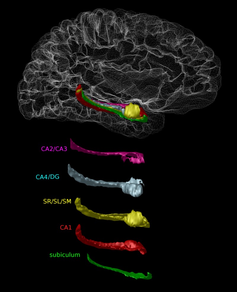
Our group has done significant work in the development of novel automated techniques for mapping the hippocampus and its subfields (Winterburn et al., NeuroImage, 2013; Pipitone et al., NeuroImage, 2014). We have developed a suite of detailed atlases that demonstrate the anatomy and architecture of this morphologically complex region of the brain. This work is currently being used to study healthy ageing and the hippocampus in the context of different neuropsychiatric disorders (see also: atlas pages).
2. Understanding the cerebellum
For decades, neuroscientific research has simply described the cerebellum as being a region that is involved in the regulation of motor function. However, emerging research suggests that the cerebellum is far more active in brain networks that are related to cognition and other brain functions. Our goal is to better understand the anatomy of the cerebellum (Park et al., 2014) and how alterations in healthy development of this understudied brain area may be implicated in different neurodevelopmental disorders such as autism and schizophrenia and neurodegenerative disorders such as Parkinson's disease.
3. Parsing disease heterogeneity
In neuroimaging research many typically ask the question: how are these brains different from one another. However, given the development and dissemination of large publicly available datasets, we are now able to ask more complex questions about the nature of neuropsychiatric disorders and their neuroanatomical signatures. However, most of these disorders are exceptionally heterogeneous in their symptomatology and in their pathological burden. Our goal is to better understand the relationship between this heterogeneity in order to better elucidate factors that are associated with risk and resilience to neuropsychiatric disorders. Projects under this theme are currently ongoing in the study of Alzheimer's and Parkinson's disease.
4. How does deep brain stimulation work?
For decades, deep brain stimulation, a therapy that involves the surgical implantation of an electrode that provides electrical stimulation to deficient brain circuits has been used in the treatment of Parkinson's disease. However, prior to the wide-spread usage of this therapy it was heavily experimented in preclinical experiments. Our goal is to better understand the brain plasticity mediated by deep brain stimulation (in clinical and preclinical experiments) that may make it an effective treatment in other neuropsychiatric disorders.
5. The shape of it all. Understanding how the shape of neuroanatomical structures is relevant and implicated in healthy development and neuropsychiatric disorders
For decades neuroimaging scientists have been using brain volume as derived from magnetic resonance imaging data as a proxy for the integrity of a myriad of neuroanatomical structures. However, we have recently developed novel and sophisticated brain mapping techniques that estimate shape indices in different structures and we demonstrate how shape is changing in the brain in the absence of volume changes; this is consistent in our studies of normative development (Raznahan et al., 2014), neurodevelopmental disorders (Shaw et al., 2014), and addiction (Janes et al., 2014). We believe that these methods can elucidate subtle brain difference that may be invisible to more traditional volumetric measures.
6. Multivariate methods for disease classification
Traditionally, scientists have used magnetic resonance imaging techniques to better quantify brain difference and neuroanatomical trends in healthy controls and disease groups. However, advances in machine learning algorithms that can handle vast quantities of data can allow us to use these data to automatically classify individuals into specific groups based solely on the neuroanatomical features that we derive. Our goal is build diagnosis algorithms that can perform the classification of individuals into disease groups based only on the information (using both native and derived measures) contained in magnetic resonance imaging and computationally derived indices of brain anatomy. We are currently developing projects that perform this type of classification in both schizophrenia and Alzheimer's disease.